Home » Without Label » Anatomy Muscles Pelvis - Pelvic Floor Anatomy Springerlink / The muscles of the pelvic floor are collectively referred to as the levator ani and coccygeus muscles.
Anatomy Muscles Pelvis - Pelvic Floor Anatomy Springerlink / The muscles of the pelvic floor are collectively referred to as the levator ani and coccygeus muscles.
Anatomy Muscles Pelvis - Pelvic Floor Anatomy Springerlink / The muscles of the pelvic floor are collectively referred to as the levator ani and coccygeus muscles.. Cross sectional muscle anatomy of pelvis, ct pelvic muscle anatomy, mri pelvic muscle anatomy, pelvic muscle anatomy, pelvic muscular anatomy, human muscles, cross sectional muscle anatomy of pelvis, ct pelvic muscle anatomy, mri pelvic muscle anatomy, pelvic muscle anatomy, pelvic muscular anatomy. Large ligaments, tendons, and muscles around the hip joint hold the bones (ball and socket) in place and keep it from dislocating. On the posterior side they are the glutei and on the anterior side the hip muscles extending into the thighs. Learn vocabulary, terms, and more with flashcards, games, and other study tools. Knowing the anatomy of this muscle can help you make good choices in caring for an.
Large ligaments, tendons, and muscles around the hip joint hold the bones (ball and socket) in place and keep it from dislocating. The muscles of the abdomen, lower back, and pelvis are separated from those of the chest by the muscular wall of the diaphragm, the critical breathing muscle. The pelvis's frame is made up of the bones of the pelvis, which connect the axial skeleton to the femurs, and therefore acts in weight bearing of the upper body. Some of the largest and most powerful muscles in the body are the gluteal muscles or gluteal group. Describe the muscles of pelvic diaphragm.
Pelvis Hip Anatomy from uploads-ssl.webflow.com (1) the obturator internus and the piriformis, which are muscles of the lower extremity, and will be described with these (pages 476 and 477); The floor of the pelvis is made up of the muscles of the pelvis, which support its. The muscles of the abdomen, lower back, and pelvis are separated from those of the chest by the muscular wall of the diaphragm, the critical breathing muscle. The right and left hip bones also converge anteriorly to attach to each other. Each hip bone, in turn, is firmly joined to the axial skeleton via its attachment to the sacrum of the vertebral column. It can be divided into the greater pelvis and the lesser pelvis. The levator ani muscles are the largest group of muscles in the pelvis. Browse 1,348 pelvis muscles stock photos and images available, or start a new search to explore more stock photos and images.
Continence, then pelvic muscle exercises may be effective.
The pelvic floor muscles provide foundational support for the intestines and bladder. Describe the muscles of pelvic diaphragm. Some of the largest and most powerful muscles in the body are the gluteal muscles or gluteal group. The pelvis's frame is made up of the bones of the pelvis, which connect the axial skeleton to the femurs, and therefore acts in weight bearing of the upper body. Large ligaments, tendons, and muscles around the hip joint hold the bones (ball and socket) in place and keep it from dislocating. Knowing the anatomy of this muscle can help you make good choices in caring for an. On the posterior side they are the glutei and on the anterior side the hip muscles extending into the thighs. Human hip musculature, computer artwork. The floor of the pelvis is made up of the muscles of the pelvis, which support its. These muscles have attachments to the pelvis as follows: The muscles of the pelvis and hip control the vast range of movement of the legs and torso. The levator ani muscles consist of three. These muscles move the thigh toward the body's midline.
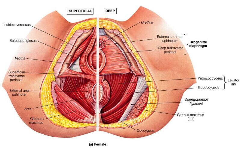
Muscles an important group of muscles in the pelvis is the pelvic floor. Arcus tendineus levator ani and the ischial spine The muscles of the abdomen, lower back, and pelvis are separated from those of the chest by the muscular wall of the diaphragm, the critical breathing muscle. (2) the levator ani and the coccygeus, which together form the pelvic diaphragm and are associated with the pelvic viscera. Human hip musculature, computer artwork.
The Pubococcygeal Muscle Pc Muscle And Attachments Yoganatomy from www.yoganatomy.com They are also known as the inner hip muscles and deep external rotators. The muscles of the pelvic floor are collectively referred to as the levator ani and coccygeus muscles. 12 photos of the muscle anatomy pelvis. The pelvic floor muscles provide foundational support for the intestines and bladder. The muscles of the pelvis and hip control the vast range of movement of the legs and torso. The psoas major and iliacus make up the iliopsoas group. On the other hand, if portions of those muscles are irretrievably lost, for example, due to complete. These are the piriformis, obturator internus, obturator externus, gemellus superior, gemellus inferior, and quadratus femoris.
Muscles an important group of muscles in the pelvis is the pelvic floor.
On the anterior side, the most prominent of the muscles are the sartorius muscle and the four muscles that make up quadriceps muscle group (the quads.) the quadriceps sounds like it should be just one muscle, akin to the triceps brachii, but it is a group of four muscles, three visible on the surface, and the fourth obscured. These are the piriformis, obturator internus, obturator externus, gemellus superior, gemellus inferior, and quadratus femoris. Small and deep muscles which mainly externally rotate the thigh at the hip joint and stabilize the pelvis. Knowing the anatomy of this muscle can help you make good choices in caring for an. (1) the obturator internus and the piriformis, which are muscles of the lower extremity, and will be described with these (pages 476 and 477); The three muscles of the group—the iliacus, the psoas major, and the psoas minor—arise from different areas of your pelvis and lumbar spine to form a common attachment in your hip. There are 10 of the 11 hip flexor muscles illustrated here. Continence, then pelvic muscle exercises may be effective. Cross sectional muscle anatomy of pelvis, ct pelvic muscle anatomy, mri pelvic muscle anatomy, pelvic muscle anatomy, pelvic muscular anatomy, human muscles, cross sectional muscle anatomy of pelvis, ct pelvic muscle anatomy, mri pelvic muscle anatomy, pelvic muscle anatomy, pelvic muscular anatomy. The pelvic girdle (hip girdle) is formed by a single bone, the hip bone or coxal bone (coxal = hip), which serves as the attachment point for each lower limb. Case contributed by assoc prof craig hacking. The adductor muscle group, also known as the groin muscles, is a group located on the medial side of the thigh. The levator ani muscles consist of three.
They form a large sheet of skeletal muscle that is thicker in some areas than in others. Some of the largest and most powerful muscles in the body are the gluteal muscles or gluteal group. The adductor muscle group, also known as the groin muscles, is a group located on the medial side of the thigh. The pelvic floor muscles provide foundational support for the intestines and bladder. It takes origin from the inner aspect of pelvis along a line extending from the body of the pubis to the ischial spine.
Hypertonic Pelvic Floor Dysfunction Vaginismus The Sexmed Advocate from images.squarespace-cdn.com It takes origin from the inner aspect of pelvis along a line extending from the body of the pubis to the ischial spine. Knowing the anatomy of this muscle can help you make good choices in caring for an. The muscles of the pelvic floor are collectively referred to as the levator ani and coccygeus muscles. Muscles that attach from the pelvis to the trunk and cross the lumbosacral joint muscles that attach from the pelvis to the thigh/leg and cross the hip joint pelvic floor muscles that are located wholly within the pelvis The pelvic floor muscles provide foundational support for the intestines and bladder. Reviews the functional anatomy of the pelvic floor structures, the effects of age on urethral support and the urethral sphincter, and attempts to clarify Included within the chart are gorgeous illustrations of the pelvic diaphragm, sphincter muscles, gluteus maximus muscles, and over a dozen more. (1) the obturator internus and the piriformis, which are muscles of the lower extremity, and will be described with these (pages 476 and 477);
Learn vocabulary, terms, and more with flashcards, games, and other study tools.
The muscles of the pelvic floor are collectively referred to as the levator ani and coccygeus muscles. These muscles move the thigh toward the body's midline. Each hip bone, in turn, is firmly joined to the axial skeleton via its attachment to the sacrum of the vertebral column. Knowing the anatomy of this muscle can help you make good choices in caring for an. Lying exposed between the protective bones of the superiorly located ribs and the inferiorly located pelvic girdle, the muscles of this region play a critical role in protecting the. Most muscles that insert on the femur (the thigh bone) and move it, originate on the pelvic girdle. Reviews the functional anatomy of the pelvic floor structures, the effects of age on urethral support and the urethral sphincter, and attempts to clarify Muscles an important group of muscles in the pelvis is the pelvic floor. Describe the muscles of pelvic diaphragm. Some of the largest and most powerful muscles in the body are the gluteal muscles or gluteal group. Learn vocabulary, terms, and more with flashcards, games, and other study tools. Large ligaments, tendons, and muscles around the hip joint hold the bones (ball and socket) in place and keep it from dislocating. The muscles of the pelvis, hip and buttock anatomical chart shows how each muscle in this area of the body works with the others, and the various minor systems within the major ones.